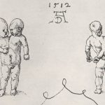
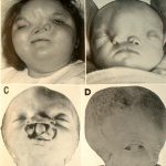
Conjoined twins (also known as Siamese twins) are a pair of identical twins born with part of their body joined together, sharing tissue which ranges from a band of flesh to major organs.

Cause
Identical twins develop when a single fertilized egg, also known as a monozygote, splits during the first two weeks of conception. Conjoined twins form when this split occurs after the first two weeks of conception. The monozygote does not fully separate and eventually develops into a conjoined fetus that shares one placenta, one amniotic sac, and one chorionic sac. Because the twins develop from a single egg, they will also be the same sex. The extent of separation and the stage at which it occurs determine the type of conjoined twin, i.e., where and how the twins will be joined.Conjoined twins originate from a single fertilized egg so they are always identical and same-sex twins. The developing embryo starts to split into identical twins within the first two weeks after conception but then stops before completion, leaving a partially separated egg which continues to mature into a conjoined fetus.
Stats
There is an extremely rare form of identical twins that occurs perhaps in one out of every 75,000 to 100,000 births or 1 in 200 deliveries of identical twins, that of conjoined twins. The birth of two connected babies can be extremely traumatic and approximately 40-60% of these births are delivered stillborn with 35% surviving just one day.
The overall survival rate of conjoined twins is somewhere between 5-25% and historical records over the past 500 years detail about 600 surviving sets of conjoined twins with more than 70% of those surviving pairs resulting in female twins.
For some reason, female siblings seem to have a better shot at survival than their male counterparts. Although more male twins conjoin in the womb than female twins, females are three times as likely as males to be born alive. Approximately 70 percent of all conjoined twins are girls.
- 73 per cent are connected at mid torso (at the chest wall or upper abdomen)
- 23 per cent at lower torso (sharing hips, legs or genitalia)
- 4 per cent at upper torso (connected at the head)
Classification of Conjoined Twins
Isidore Geoffroy Saint-Hillaire was the first teratologist to classify conjoined twins, using Greek etymology to describe the twins in terms of their shared anatomy. Many of his terms are still in use today. Listed below are some commonly occurring types of conjoined twins. All of these types can be more broadly categorized as displaying either equal and symmetrical forms or unequal and possible asymmetrical forms.
- Craniopagus Twins are joined at the cranium (the top of the head or skull). Occuring in just 2% of all conjoined twin cases, this is a very difficult type of twin to separate although advances in medicine have led to more than 35 successful separations. Two female craniopagus twins were successful separated in Lithuania in 1989, for example.
- Thoraopagus The most common form of conjoined twins, occuring in between 35-40% of all cases. The twins share part of the chest wall, possibly including sharing the heart.
- Pygopagus Twins are likely positioned back-to-back and usually have a posterior connecton at the rump. Occurs in almost 20% of documented cases.
- Ischiopagus About 6% of all conjoined twins have this condition, with the twins joined by the coccyx (lowest part of the backbone) and the sacrum (backbone immediately above the coccyx).
- Omphalopagus Twins are united from the waist to the lower breastbone, probably accounting for about 34% of conjoined cases.
- Dicephalus One body with two separate heads and necks. Abigail and Brittany Hensel of the United States are an example of this very rare type of conjoined twin. The Tocci Brothers, Scottish Brothers and Ritta and Christina were also examples of this type of conjoined twin.
Dr. Rowena Spencer’s 2003 book Conjoined Twins: Developmental Malformations and Clinical Implications discusses several cases of supposed conjoined triplets and quadruplets, most of which consist of one autosite with a number of parasites or acardiac twins (see above). However, a 2004 article in The American Journal of Obstetrics and Gynecology describes a case of parapagus dicephalus dibrachius dipus twins with triplet joined to the shared sternum, in the manner of xiphopagus twins. All three fetuses were well-formed and had approximately normal heads and extremities. This article provides conclusive proof that conjoined triplets, although extremely uncommon, can occur.
History
One of the earliest documented cases of conjoined twins were Mary and Eliza Chulkhurst. They were born in Biddenden, County of Kent, England in the year 1100, and were joined at the hip.
The wealthy sisters, who were known as the Biddenden Maids, lived for 34 years. When they died, they left a small fortune to the Church of England. In honor of their generosity, it was customary for English citizens to bake little biscuits and cakes in the sisters’ images and give them to the poor.
Another set of famous conjoined twins was Eng and Chang Bunker
The most famous set of conjoined twins were Chang and Eng, the men who originated the term "Siamese Twins". Eng and Chang were born in Siam (modern day Thailand) on May 11, 1811 to a Chinese father and half-Chinese, half-Malay mother. Thanks to their heritage, while growing up in Siam the boys were known as "The Chinese Twins".
Separation
Today, most pairs of conjoined twins are successfully identified during prenatal examinations. Some types of conjoined twins are much easier to separate while other rare forms lead to complicated and costly procedures that can lead to difficult ethical and moral decisions of separation surgery, especially if the twins share internal organs or if they are joined at the head.
Recent successful separations of Siamese twins include that of the separation of Ganga & Jamuna Shreshta in 2001, who were born in Kathmandu, Nepal in 2000. The 97 hour surgery on the pair of craniopagus twins was a landmark one which took place in Singapore; the team was led by neurosurgeons, Dr. Chumpon Chan and Dr. Keith Goh.
In 2003 two women from Iran, Ladan and Laleh Bijani, who were joined at the head but had separate brains (craniopagus) were surgically separated in Singapore, despite surgeons’ warnings that the operation could be fatal to one or both. Both women died during surgery on July 8, 2003.
According to the book ‘Entwined Lives’, there have been approximately 200 attempted surgical separations of conjoined twins, with 90% of these occurring after 1950. Three-quarters of the procedures since 1950 have resulted in one or both of the twins surviving.
Several hospitals across the world specialize in these often difficult surgeries. The Children’s Hospital of Philadelphia has performed more than a dozen successful separation surgeries since its first operation in 1957 (most recently performing one on 7-month-old twin girls on March 1, 2001), while physicians such as Dr. Rich Hampton of the Pediatric Surgical Associates in the Twin Cities of Minneapolis and St. Paul, successful separated Keri and Kaci Archer in October, 1991. The Children’s Hospital of Boston has operated on nine sets of conjoined twins (as of the mid 1990’s, current numbers not available).
There are organizations which can help parents of conjoined twins address some of the issues they will face during their pregnancies or while raising their children. Conjoined Twins International, based in Prescott, Arizona, was founded in 1987 by a grandfather of conjoined twins. The organization gives advice and support to a little more than half of the families of conjoined twins currently living in the United States.


